KAIST
BREAKTHROUGHS
Research Webzine of the KAIST College of Engineering since 2014
Spring 2025 Vol. 24Successful development of a simultaneously PET-MRI imaging system
A simultaneously PET-MRI imaging system was successfully developed based on new SiPM detectors.
Article | Spring 2014
Expectations of the localization of medical imaging technology are increasing. A research team consisting of members from KAIST, Seokang University, and Seoul National University with the collaboration of KAIST Nanofabrication Center and led by Professor Gyuseong Cho from the Nuclear and Quantum Engineering Department of KAIST, has successfully developed a simultaneous PET-MRI imaging system using indigenous domestic technology and obtained sample brain images from three different volunteers.
PET-MRI is a medical imaging system that is considered to be the next-generation of multi-modality imaging techniques. The system is composed of Positron Emission Tomography (PET), which can diagnosis the function of the human body and Magnetic Resonance Imaging (MRI), which can investigate the anatomy of the human body. Since the combination of two different modalities into a single system gives an advantage in investigating both the anatomical and functional activity of our body simultaneously, it is possible to precisely diagnosis and treat diseases that were difficult to identify in the past, e.g., various cancers, Alzheimer’s, Parkinson’s, and other brain-related injuries.
However, existing devices have to acquire PET and MRI images separately due to the high magnetic field from the MRI, thus leading to longer shooting time and more error due to the patient’s movement. Because of this, it is necessary to move towards a system that can gain both images together.
The research team mainly focused on three areas: PET detectors that get no interference from magnetic fields, PET-MRI fusion systems, PET-MRI image processing.
PET detectors are the core components that take up half the manufacturing cost of PET systems. KAIST Nuclear and Quantum Engineering Professor Gyu Seong Cho and National NanoFab Center Researcher Wu Seok Seol, along with their research teams, have successfully developed a silicon Photo Multiplier Tube that can be used in magnetic fields with domestic technology. The newly developed sensor has gained global competitiveness thanks to a mass production rate of 95% using an optimized semiconductor process and a gamma energy resolution in the 10% range.
Based on these technologies, in June, the joint research team successfully developed a brain PET-MRI system and obtained three fusion images. The team reported that these were the first human images in the world from a silicon PMT-based PET and MRI joint system.
A major advantage of this system is that it can acquire a PET-MRI image with a low installation cost due to the detachable brain PET module and MRI head coil on the existing whole-body MRI system.
Professor Cho explained, “With this successful development, we were able to provide a standard for domestically-produced commercial PET systems and stand shoulder to shoulder with other private enterprises. In addition, we project the demand for PET-MRI system will increase in medical fields, particularly those related to brain injuries such as Alzheimer’s and Parkinson’s because this new state-of-the-art medical imaging system is excellent in cost and performance.”


Most Popular

When and why do graph neural networks become powerful?
Read more
Smart Warnings: LLM-enabled personalized driver assistance
Read more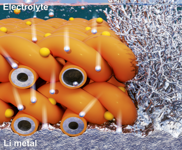
Extending the lifespan of next-generation lithium metal batteries with water
Read more
Professor Ki-Uk Kyung’s research team develops soft shape-morphing actuator capable of rapid 3D transformations
Read more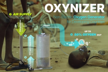
Oxynizer: Non-electric oxygen generator for developing countries
Read more