KAIST
BREAKTHROUGHS
Research Webzine of the KAIST College of Engineering since 2014
Spring 2025 Vol. 24Visualization of 3D microscopic structures of blood vessels using an ultra-high-speed intravascular endoscopic microscopy system
Visualization of 3D microscopic structures of blood vessels using an ultra-high-speed intravascular endoscopic microscopy system
Ultrahigh-speed endoscopic optical coherence tomography (OCT) system visualizes 3D microscopic structure of the inside of blood vessels.
Article | Fall 2014
Heart attack, most frequently caused from the failure of the coronary artery of the heart, which supplies blood to the heart muscles, is the leading cause of death in industrialized countries. The recent development of high-speed second-generation (2G) Optical Coherence Tomography (OCT) enables endoscopic microscopy of patients’ long coronary artery segments, which opens up new horizons in diagnostics of high-risk coronary plaques and will hopefully lead to the development of new therapeutic methods.
Although OCT is the only imaging method that can provide sufficient resolution to visualize important microscopic features associated with high-risk coronary lesions, true high-resolution 3D visualization has not been achieved for long coronary segments because longitudinal spatial resolution is several times poorer than the cross-sectional resolution due to still insufficient imaging speed.
Prof. Wang-Yuhl Oh’s research group recently developed an ultra-high-speed intravascular OCT (IV-OCT) system and demonstrated the first true high-resolution 3D visualization of the microstructure of the vessel wall in vivo. Development of a high-speed 2G-OCT system, a miniaturized optical catheter (endoscopic probe), and a high-speed and high-precision fiber-optic rotary junction enables high-speed IV-OCT imaging that is 3-8 times faster than the current fastest IV-OCT.
Images of rabbit aorta obtained by the developed high-speed IV-OCT system (Figures 1 (a), (b), and (d)) visualize the microstructures of the vessel, such as the stent strut microstructure and the orifice of the side branch, much better than the ones imaged with the currently fastest speed (Figures 1 (c) and (e)). If the developed system images with the conventional longitudinal pitch, it only takes 1 second to image a 7.2cm-long segment of vessel.
Prof. Oh’s group currently is in the process of in-vivo coronary artery imaging of large animals, such as swine, and is planning to conduct clinical patients’ coronary artery imaging afterward.
This work was supported in part by the NRF of Korea, grant 2010-0017465 and by the MSIP of Korea, grant GFP/(CISS-2012M3A6A6054200), and was published on Jan. 1, 2014 in Biomed Opt Express.

Most Popular

When and why do graph neural networks become powerful?
Read more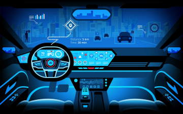
Smart Warnings: LLM-enabled personalized driver assistance
Read more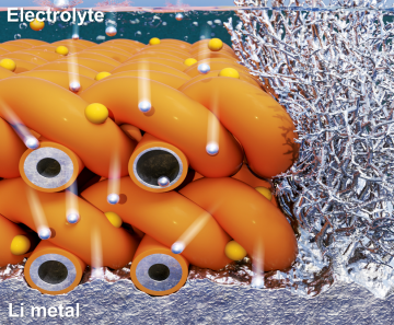
Extending the lifespan of next-generation lithium metal batteries with water
Read more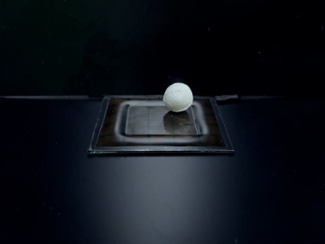
Professor Ki-Uk Kyung’s research team develops soft shape-morphing actuator capable of rapid 3D transformations
Read more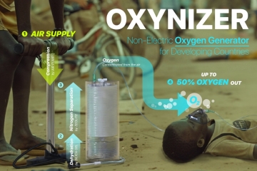
Oxynizer: Non-electric oxygen generator for developing countries
Read more