KAIST
BREAKTHROUGHS
Research Webzine of the KAIST College of Engineering since 2014
Spring 2025 Vol. 24Never-fading pigments developed by colloidal crystals confined in microcapsules
Never-fading pigments developed by colloidal crystals confined in microcapsules
Uniform colloids are assembled to form a crystalline phase in the internal volume of microcapsules, exhibiting sparkling structural colors through optical interference. The colors developed by a regular lattice of colloids never fade as long as the structure is maintained, and the lattice structure can be further engineered to modulate the colors through external fields.
Article | Fall 2014
Colors are usually developed by chemical pigments, which absorb specific wavelengths of visible light. For example, the green color of leaves is made by chlorophyll, which absorbs blue and red bands of visible light. Such chemical colors fade as the chemical structure is transformed by external stimuli such as ultraviolet. Unlike chemical colors, structural colors are developed by regular nanostructures that provide constructive interference of selected wavelengths. Therefore, structural colors never fade as long as the structures remain intact.
Nature has developed such structural colors over a long evolutionary period. Morpho butterflies, birds of paradise, and emperor penguins are limited examples of animals that have structural colors on their bodies. Humans have recently tried to develop structural colors by creating regular nanostructures with the assistance of self-assembly; however, humans have been developing such colors for less than 30 years, while nature has developed them for millions of years. One of the most practical approaches developed by humans is colloidal self-assembly.
Uniform colloids spontaneously form crystalline phase when they are concentrated. The colloidal crystals reflect a selected wavelength of light through constructive interference, and structural colors are developed when the periodicity is half the wavelength of visible light. Typically, colloidal photonic structures are prepared in the form of thin solid film. However, such colloidal crystals are not reconfigurable or mechanically stable, thereby severely restricting further processing.
To overcome these limitations, Prof. Shin-Hyun Kim’s group developed photonic microcapsules in collaboration with the Prof. Daivd Weitz and Prof. Vinnothan Manoharan groups in Harvard University. With a capillary microfluidic device, aqueous colloidal suspension is encapsulated with an ultra-thin oil shell in continuous water phase, forming core-shell structures; this is referred to as double-emulsion drops. The colloids in the core are concentrated by the selective removal of water through the thin shell by positive osmotic pressure (Figure 1). This leads to the crystallization of colloids, and therefore, structural color is developed in each microcapsule. The oil shell is photo-polymerized to provide a solid membrane with tunable rigidity.
The ink capsule of the colloidal photonic structure provides ease of processing and high reconfigurability (Figure 2). Therefore, the capsules can be employed as building blocks to construct photonic devices. Furthermore, colloids confined in each microcapsule remain mobile as they are dispersed in a liquid core, which allows rapid modulation of the structural colors through external fields, making the photonic capsules more valuable for color displays operated at reflection mode.
An article on this research (entitled “Osmotic-pressure-controlled concentration of colloidal particles in thin-shelled capsules”) was published on January 7, 2014 in Nature Communications.
Reference: S.-H. Kim. Osmotic-pressure-controlled concentration of colloidal particles in thin-shelled capsules. Nat. Commun. 5:3068 (2014)
Additional link for more information:
http://www.nature.com/ncomms/2014/140107/ncomms4068/full/ncomms4068.html


Most Popular

When and why do graph neural networks become powerful?
Read more
Smart Warnings: LLM-enabled personalized driver assistance
Read more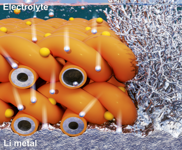
Extending the lifespan of next-generation lithium metal batteries with water
Read more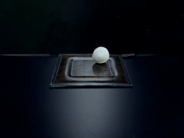
Professor Ki-Uk Kyung’s research team develops soft shape-morphing actuator capable of rapid 3D transformations
Read more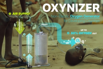
Oxynizer: Non-electric oxygen generator for developing countries
Read more