KAIST
BREAKTHROUGHS
Research Webzine of the KAIST College of Engineering since 2014
Spring 2025 Vol. 24Non-thermosensitive cell-sheet engineering via functional polymer coatings
Non-thermosensitive cell-sheet engineering via functional polymer coatings
Prof. Sung Gap Im’s group developed a new cell sheet technology that can harvest cell sheets within 100 seconds without changing temperature through a functional polymer film that can precisely control the surface free energy of the culture substrate.
Article | Fall 2020
Prof. Sung Gap Im’s group reported a new cell sheet technology that can harvest cell sheets within 100 seconds without changing the temperature. The study appears in the March 2020 edition of Advanced Materials under the title, “A Surface-Tailoring Method for Rapid Non-thermosensitive Cell Sheet Engineering via Functional Polymer Coatings .”
If the skin, cartilage, nerves, myocardial tissues, or other parts of the body are severely damaged, tissue may need to be transplanted. Despite the enormous demand, the amount of tissue that can be transplanted is extremely limited. Therefore, the restoration and regeneration of living tissues necessitates the development of a treatment for regenerating tissue from a patient’s cells into a tissue capable of living transplantation. In order to achieve excellent tissue regeneration, “cell sheet” engineering that allows cell detachment from the substrate without damage.
Prof. Im’s group has developed a new surface modification technology that can harvest cell sheets in a short time (100 seconds) without changing temperature through a functional polymer thin film that can precisely control the surface free energy of the culture substrate. Through this, it was confirmed that the formation of various kinds of cell sheets was possible, and the cell damage was greatly reduced, thereby demonstrating that the method is a highly versatile technology capable of recovering cell sheets with high survival rate without cell cycle and DNA loss.

In addition, when the recovered cell sheets were stacked in multiple layers and transplanted into diabetic ulcer and ischemic rats, the treatment efficiency was significantly improved compared to the conventional method. In particular, this method can easily harvest the cells in the form of a sheet via modulation of the adhesion strength between cells and culture surface only by replacing the cell culture medium.
The fabrication method of a novel cell sheet is entirely different from the method using temperature change; therefore, the technology is expected to contribute significantly to the field of regenerative medicine in the future.
Eunjung Lee, a research professor at KAIST who participated as corresponding author, explained, “This is an innovative cell sheet manufacturing method that can improve the graft survival rate by minimizing cell damage; thus, as a core technology for developing cell therapy products, it is expected to serve a broad range of applications.”
This research was supported by the Ministry of Science and ICT and Korea Research Foundation.


Most Popular

When and why do graph neural networks become powerful?
Read more
Smart Warnings: LLM-enabled personalized driver assistance
Read more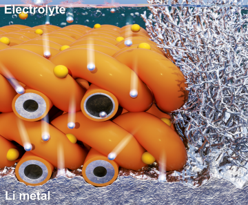
Extending the lifespan of next-generation lithium metal batteries with water
Read more
Professor Ki-Uk Kyung’s research team develops soft shape-morphing actuator capable of rapid 3D transformations
Read more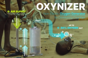
Oxynizer: Non-electric oxygen generator for developing countries
Read more