KAIST
BREAKTHROUGHS
Research Webzine of the KAIST College of Engineering since 2014
Spring 2025 Vol. 243D visualization and quantification of bioplastic PHA in living bacteria
3D visualization and quantification of bioplastic PHA in living bacteria
A KAIST research team has used 3D holographic microscopy to observe how bioplastic ‘polyhydroxyalkanoate’ (PHA) granules accumulate in living bacteria cells. 3D imaging and quantitative analysis of the PHA granules provides insight into biosynthesizing sustainable substitutes for petroleum-based plastics.
Article | Spring 2022
The bio-degradable polyester polyhydroxyalkanoate (PHA) is being touted as an eco-friendly bioplastic to replace existing synthetic plastics. While having properties similar to those of general-purpose plastics such as polyethylene and polypropylene, PHA can be used in various industrial applications such as container packaging and disposable products.
PHA is a natural polymer synthesized by numerous bacteria as an energy and carbon storage material and exists in the form of insoluble granules within cells. Previous in vivo investigations of PHA granules have been performed using fluorescence microscopy, transmission electron microscopy (TEM), and electron cryotomography. These techniques have generally relied on statistical analysis of multiple 2D snapshots of fixed cells or short-time monitoring of cells. For TEM analysis, cells need to be fixed and sectioned, and thus the investigation of living cells has not been possible. Fluorescence-based techniques require fluorescence labeling or dye staining. Thus, indirect imaging with using reporter proteins cannot show the native state of PHAs or cells, and invasive exogenous dyes can affect physiology and viability of cells. These technical limitations have made it difficult to fully understand the formation of PHA granules in cells, and thus only several mechanistic models based on observations have been proposed.
A team of metabolic engineering researchers led by Distinguished Professor Sang Yup Lee, as well as Physics Professor YongKeun Park, who established the startup Tomocube with his technology of 3D holographic microscopy, reported 3D quantitative analysis of PHA granules in live bacterial cells, performed using optical diffraction tomography to measure refractive index distributions. The formation and growth of PHA granules in cells of Cupriavidus necator, the most-studied native PHA producer, and recombinant Escherichia coli, harboring the C. necator PHA biosynthesis pathway, were intensively examined.
From the reconstructed 3D refractive index distribution of the cells, the team succeeded in 3D visualization and quantitative analysis of cells and intracellular PHA granules at single-cell level. In particular, the team newly presented the concept of “in vivo PHA granule density.” Through statistical analysis of hundreds of single cells, distinctive differences of density and localization of PHA granules in the two micro-organisms were found. Furthermore, the team identified the key protein that enables the characteristics of PHA granules in recombinant E. coli to become similar to those of C. necator.
The research team also presented 3D time-lapse movies showing the actual processes of PHA granule formation and cell growth. Living cells synthesizing and accumulating PHA granules in their native state had previously never been directly visualized.
The research team believes that a deeper understanding of PHA granule formation within bacterial cells is now possible; this study will help develop various bioplastic production processes in the future.
The research paper titled “Three-dimensional label-free visualization and quantification of polyhydroxyalkanoates in individual bacterial cell in its native state” was published in PNAS on July 26, 2021.
Most Popular

When and why do graph neural networks become powerful?
Read more
Smart Warnings: LLM-enabled personalized driver assistance
Read more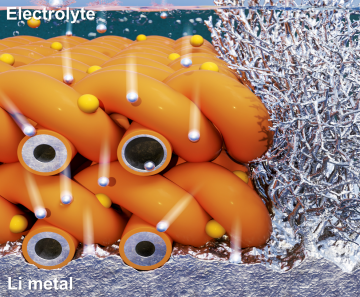
Extending the lifespan of next-generation lithium metal batteries with water
Read more
Professor Ki-Uk Kyung’s research team develops soft shape-morphing actuator capable of rapid 3D transformations
Read more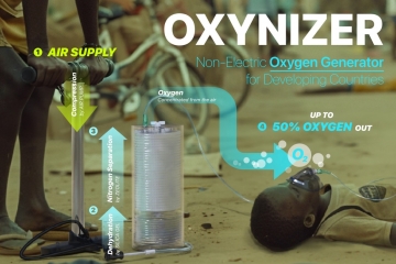
Oxynizer: Non-electric oxygen generator for developing countries
Read more